Development of three-dimensional lacrimal gland tissue in vitro
Contact
Director of the Department of Ophthalmology
Head of the Laboratory for Experimental Ophthalmology
Office at Pius-Hospital Oldenburg
Address
Development of three-dimensional lacrimal gland tissue in vitro
Lacrimal gland insufficiency is one of the main causes of the development of dry eye syndrome (keratoconjunctivitis sicca, DES), which can lead to pain, impaired vision and contrast vision and, in the most severe cases, complete loss of vision.
One of the main causes of DES is dysfunction of the lacrimal gland (TD). This can be age-related, but can also be triggered by chronic autoimmune diseases such as Sjögren's syndrome. Sjögren's syndrome manifests itself in a morphological change in the lacrimal and salivary glands, which are affected by inflammatory processes. In this case, tear fluid is either not produced at all or only to a very limited extent, or the composition of the aqueous phase of the tear film is severely altered. The worldwide prevalence of dry eye syndrome is estimated at five to 35 per cent, depending on the age group (21 - >65 years). To date, there are no successful curative treatment options, which means that, in addition to the intensive use of tear substitutes, patients are left with the use of specially made liquid chamber glasses or closure of the tear puncta by means of so-called "punctum plugs" or sclerotherapy. However, in severe cases of dry eye, these measures rarely lead to lasting relief from symptoms.
A new and promising therapeutic approach is the reconstruction of a three-dimensional, functionally active lacrimal gland (TD) using so-called "tissue engineering" methods, which is the basis of this project.
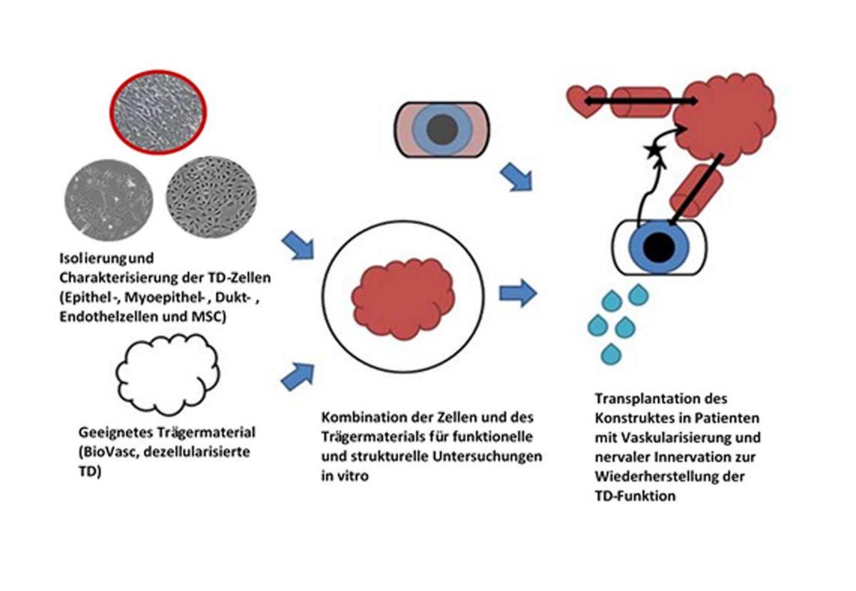
The basis of this construct is a cell-free tissue (so-called BioVasc (parts of the intestine) and lacrimal gland) from domestic pigs (sus scrofa). In addition to the good availability and the already clinically proven biocompatibility of porcine tissue (for example, the heart valve replacement tissue from pigs that has been used for years), this cell-free matrix offers the structurally necessary conditions, such as the presence of vascular and surface structure, which are necessary for the successful reconstruction of a TD. In addition to a suitable carrier matrix, the exact composition of the cell population is of decisive importance for the reconstruction of a functionally active TD. In a lacrimal gland, the functionally active acinar and ductal cells (epithelial cells) and the mesenchymal stem cells are found alongside the vascularising endothelial cells.
As part of the project, the different cell types were isolated from porcine lacrimal glands, cultured and analysed for their secretory activity in cell culture. Subsequently, organoid-like structures (so-called spheroids) were generated by using all three cell types and applied to decellularised porcine carrier material. Under dynamic culture conditions in a bioreactor, the spheroids actively secreted various substances found in the tear fluid. Following these successful initial experiments, the complexity of the 3D structure is to be adapted to the structure of a lacrimal gland in this project by utilising a BioVasc® system (and the existing vascular system).
The main objective of the study is to investigate the extent to which vascularisation of the three-dimensional carrier material influences the ingrowth and functionality of the lacrimal gland cells introduced. The different systems are analysed by means of immunohistological and functional examinations. Both the morphology of the constructs and the secretion capacity are analysed. These approaches form the basis for planned in vivo experiments in which functionally active and vascularised constructs are transplanted into animals to replace a damaged lacrimal gland. This represents a potential new therapeutic option for dry eye syndrome.